What are Rare Vascular Tumors?
Vascular birthmarks are a broad term that encompasses any abnormal development of blood vessels in the skin or internal organs. These conditions can range from mild to severe, depending on the individual case. Vascular anomalies are usually present at birth and may be present for life, though they can often be managed with treatments such as laser therapy or medical interventions
Types of Vascular Birthmarks
The most common type of vascular birthmark is known as a port-wine stain or capillary malformation. This condition appears as a flat, red-purple mark on the skin that is caused by an overgrowth of dilated capillaries, which are small blood vessels located just below the surface of the skin. Port-wine stains can occur anywhere on the body but are most commonly found on the face, neck, arms, legs, and trunk. They tend to appear darker in color when exposed to cold temperatures.
Vascular lesions can also develop internally in organs such as the liver or brain and can have more serious consequences than those seen externally in port-wine stains or hemangiomas. Diagnosis of these conditions requires an extensive physical exam as well as imaging studies such as CT scans and MRIs to properly assess their size and location within the body’s structures. Treatment for these conditions depends upon their severity; some require no intervention, while others may need surgical removal or radiation therapy if medical treatment has proven ineffective.
Regardless of what type of vascular anomaly is present, it is important for patients to understand that treatment options exist that can help reduce their appearance or alleviate any underlying symptoms they may be experiencing due to changes in endothelial cells – cells that line our blood vessels – caused by these debilitating conditions
Call To Action
Call us today!
Infantile Hemangiomas
Infantile Hemangiomas are a type of congenital or birthmark vascular malformation that affects many pediatric patients. These benign tumors can appear anywhere on the body and are commonly seen in the head, neck, upper trunk, and extremities. They usually become apparent within the first few weeks after birth but can occasionally be present at birth. While most cases of infantile hemangiomas will resolve without treatment by 16 to 18 months of age, some require medical intervention to ensure proper healing and reduce potential risks such as bleeding or ulcerations.
Superficial capillary hemangiomas are the most common type of infantile hemangioma and typically appear as flat red-purple skin lesions that spread out from the central area. These lesions occur due to an abnormal growth of blood vessels in the affected area, which leads to increased blood flow to this region. If not treated, superficial capillary hemangiomas may increase in size over several months or years before eventually resolving on their own.
Epithelioid Hemangioendothelioma is a rarer form of infantile hemangioma that is seen more often in older children and adults than infants and young children. This type of vascular tumor appears as a soft tissue mass and can cause significant changes in blood pressure if not treated quickly with interventional radiology procedures or plastic surgery techniques such as laser treatment or dye laser treatment. Genetic testing may sometimes be used to identify any underlying risk factors for developing this type of tumor, although this is not always possible due to its rarity among pediatric patients.
Overall, about 75 percent of all infantile hemangiomas typically resolve within a few months up to two years after they first appear; however, depending upon their location and severity, some may require medical intervention for proper resolution. It is important for parents to monitor their child’s growth closely so that any changes in size or color can be reported promptly to the child’s healthcare provider so that appropriate treatments can be recommended if necessary
Hemangiomas in Skin Diagnosis
As an expert plastic surgeon, hemangiomas in skin diagnosis is a complex topic that requires careful evaluation of a patient’s medical history, physical examination, and laboratory tests to ensure correct diagnosis. Hemangiomas are vascular tumors that can form anywhere on the body but most commonly appear on the skin and can be either superficial or deep. Superficial lesions affect only the top layer of the skin and usually resolve without treatment, whereas deep hemangiomas have deeper components that may involve vital organs or lymphatic vessels as well as the skin.
In order to make an accurate diagnosis, it is important for physicians to consider both physical examination findings and appropriate laboratory tests, such as blood tests, to assess any underlying health issues that could contribute to the development of this condition. Additionally, prospective studies using imaging techniques such as CT scans or MRIs can be used to determine if there are any deep components present and how extensive they might be. Once all of this information has been gathered, a comprehensive clinical study should be conducted in order to determine what type of standard treatment should be applied in order to achieve satisfactory results.
Overall, hemangioma diagnosis is a complex process made up of multiple elements that must all work together in order for a correct diagnosis and successful treatment plan to be developed. It is essential for healthcare providers to take into account both shallow and deep components when making their assessment so that any potential risks associated with these lesions can be properly managed before they cause further damage or complications.
How to Treat Vascular Birthmarks
The key to successful treatment of vascular birthmarks is early detection and diagnosis. It is important for healthcare professionals to detect and diagnose any potential vascular birthmarks as soon as possible in order to ensure proper management and prevent further complications from occurring. Depending on the type, size, and location of the birthmark, treatment may involve interventional radiology techniques such as laser therapy or microsurgery. Laser therapy can be used to reduce the amount of blood flow to the lesion, while microsurgery can be used to excise or remove the tissue that has been affected by the birthmark.
In more severe cases where the lesion is large or located in an area that cannot be easily treated with lasers, chemotherapy may be recommended in order to shrink or stop the growth of the abnormal tissue. Additionally, there are medications available that have been shown to decrease inflammation associated with certain types of vascular birthmarks, which can help minimize scarring and improve overall aesthetic outcomes. In some cases, medical treatments can be combined with plastic surgery techniques such as skin grafting or reconstructive surgery in order to achieve optimal results.
No matter what type of treatment is being considered for a patient’s vascular birthmark, it is essential for healthcare providers to provide comprehensive education about potential risks and benefits as well as give clear instructions about how best to manage their condition over time. By taking all these steps into consideration during a targeted treatment plan, physicians can ensure that patients receive effective care that will result in improved quality of life and long-term health benefits associated with properly managed vascular birthmarks.
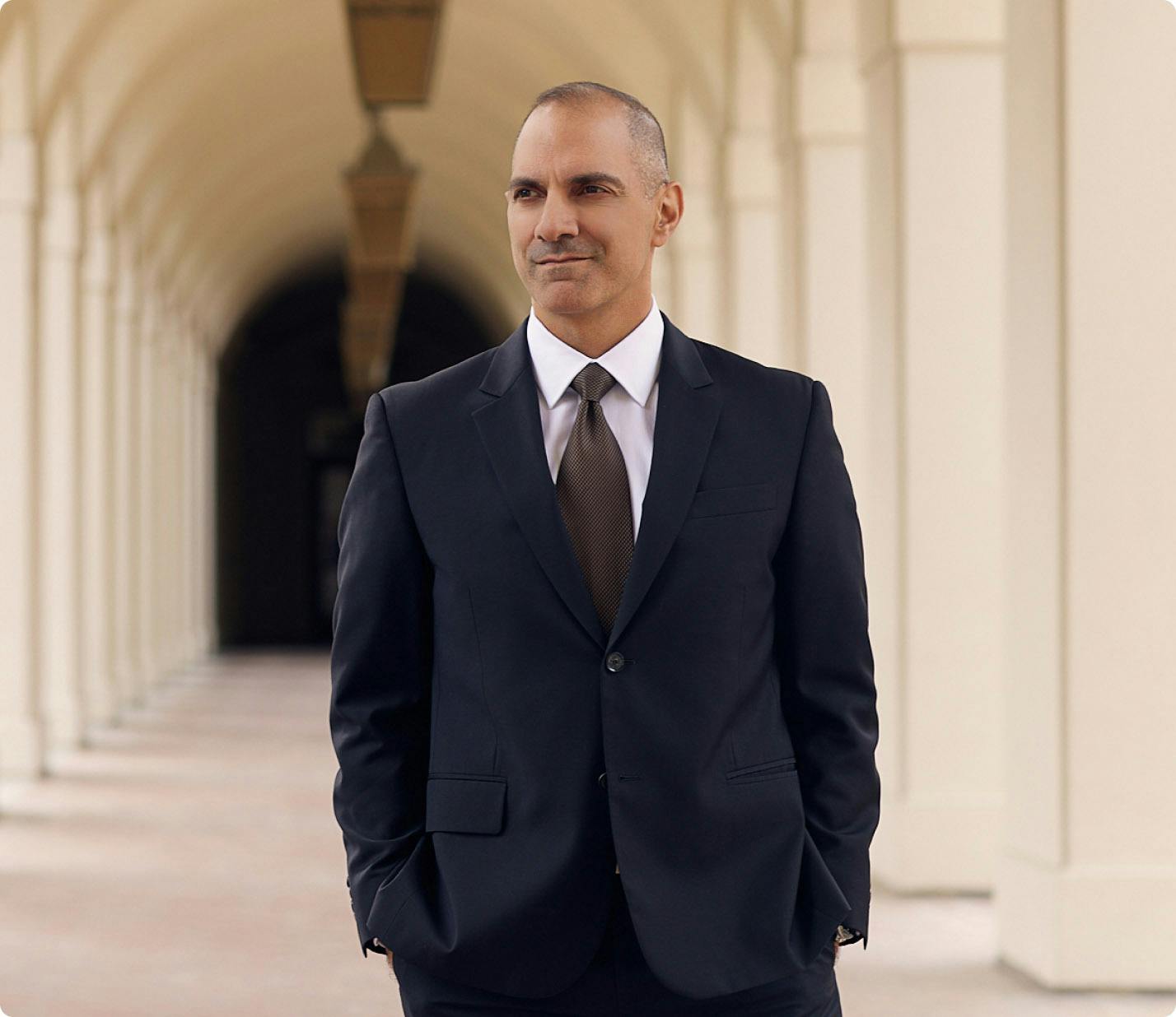
Why choose Dr Panossian
- He received his medical education at Tufts University School of Medicine.
- Graduated at the top of his class at UCLA, receiving Phi Beta Kappa and Summa Cum Laude honors.
- Was accepted into an elite combined general surgery and plastic surgery residency at the Keck School of Medicine of USC.
- Completed subspecialty training in craniofacial surgery at the Hospital for Sick Children in Toronto and Harvard Medical School.
- Was mentored by Dr. Ron Zuker in the practice of facial paralysis reconstruction. This prestigious fellowship position was available to only one surgeon in the United States.
- Is affiliated with various charitable and educational organizations, including Operation Smile and Mending Kids.
- Is a member of the American Society of Plastic Surgeons and the highly selective American Association of Plastic Surgeons, reserved for only a select group of individuals nationally who have demonstrated excellence in academic plastic surgery.
- Holds memberships in several other professional societies including the American College of Surgeons and the American Society of Reconstructive Microsurgery.
- He serves on the Board of Directors for Mending Kids and the Gondobay Manga Foundation, a non-profit organization dedicated to the improvement of lives in Sierra Leone.
- Has been nominated by his peers annually since 2012 as a “Super Doctor.”
- Served as an expert medical consultant and appeared on The Doctors, Grey’s Anatomy, and Nip/Tuck.
- Has been featured as “Top Doctor” in US News and World Report, Pasadena Magazine, and Los Angeles Magazine.
Last modified by Dr. Andre Panossian