Gracilis Muscle Transplant (Cross Face Nerve Graft)

What is a gracilis muscle transplant with cross-face nerve graft?

The gracilis muscle transplant with cross-face nerve graft consists of two surgeries to reanimate the smile. Prior to harvesting the gracilis muscle, an initial surgery is required to bring innervation from the unparalyzed side of the face. This is known as a cross-face nerve graft.

(626) 765-6885

Girl with facial paralysis before and after cross face nerve graft and gracilis muscle transplant.

During this initial surgery, a long nerve is harvested from the lower leg (sural nerve). This nerve supplies only a small patch of sensation to the outer part of the foot, and therefore, results in no noticeable loss of function. It is typically harvested from the leg with three small incisions and does not result in disfigurement. The nerve is then implanted into the unparalyzed side of the face to one of the working branches of the facial nerve responsible for generating an ideal smile. It is then tunneled within the face and banked in the upper lip, close to the paralyzed side of the face. After a period of 6-12 months, nerve regeneration reaches the terminal point of the nerve graft and is ready for powering the gracilis muscle.

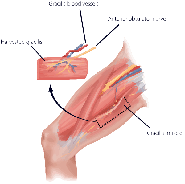
The gracilis is a long muscle located in the inner thigh. It is responsible for moving the hip inwards (or, adduction). It is a redundant muscle for this function. In other words, there are several other muscles that accomplish the same goal, therefore, sacrificing the muscle results in no loss of function in the thigh. Only a small, thin segment of the gracilis muscle is taken from the thigh along with its blood and nerve supply. The muscle is then moved to the paralyzed side of the face where it is connected to local nerves and blood vessels. In this case, it is attached to the cross-face nerve graft. After a period of 6-12 months, the muscle becomes active and contracts to move the corner of the lip.

The primary advantage of using a cross-face nerve graft to power the gracilis muscle is the spontaneous nature of the smile following reanimation. In other words, the neural pathways for initiating a smile occur through normal channels via the facial nerve. Therefore, there is no re-training of muscles required as there would be with temporalis muscle transfers or gracilis muscles connected to the masseter nerve (fifth cranial nerve used for biting).

How is a gracilis muscle transplant with cross-face nerve graft performed?

For details on the cross-face nerve graft, visit the cross-face nerve graft section. The gracilis muscle is harvested through a small incision measuring 3-4 inches in the inner thigh. The blood vessels and nerves are isolated. The muscle is measured for the exact length and width that is required in the paralyzed side of the face, from the corner of the lip to an area near the top of the ear. Once the dimensions are confirmed, the muscle is then split and divided. It is then transplanted to the face where it is anchored. In preparing the face, an incision is placed in the pattern of a facelift with a small extension underneath the jaw. The skin is elevated, and anchoring sutures are placed at precise locations along the corner of the mouth and upper lip. The blood vessels and the previously placed cross-face nerve graft are located and prepared. The blood vessels are then connected using a high-powered microscope (microsurgery). Next, the nerve is connected in the upper lip to the cross-face nerve graft. All incisions are closed with absorbable sutures.

Diagram of gracilis muscle transplant for facial paralysis reconstruction.
Diagram of gracilis muscle
transplant for facial
paralysis reconstruction.

What is the recovery from a gracilis muscle transplant with cross-face nerve graft?

For recovery following a cross-face nerve graft, refer to the appropriate section. Following the second surgery for the gracilis muscle transplant, patients remain in the hospital or aftercare facility for 3-6 days. The head is kept wrapped in a bulky soft dressing, and the thigh is wrapped with a mild compression dressing. There may be moderate swelling of the face and eyelids. Antibiotics are required for 10-14 days, and a soft diet is necessary for 3 weeks. A soft rubber drainage tube is left underneath the skin to relieve any fluid collection in the face. This is taken out 2-3 days following surgery. It is imperative to avoid any pressure or accidental blunt trauma to the cheek during this time. Contact sports and heavy lifting are avoided for 4-6 weeks. Adults will need to take between 3-4 weeks off for recovery before they return to light duty. Muscle activity then begins approximately 3-9 months after surgery.

I had a very professional experience for both of my surgeries.

  • Star full Star full Star full Star full Star full 5 stars
  • Leon Armstrong

View all Testimonials

What are the risks and complications of a gracilis muscle transplant with cross-face nerve graft?

Bleeding, infection, and complications from anesthesia are possible during or after surgery, as with all types of surgical procedures. These are rare risks; however, other risks are related more specifically. For example, asymmetry is possible either in the resting position or with smiling. The amount of muscle movement is difficult to predict with accuracy at the time of surgery. Also, there may be contour issues such as increased cheek bulk or unwanted dimpling of the cheek with smiling. The creation of the lip-cheek fold (nasolabial fold) may also be unpredictable. In addition, there is a small rate of failure that may be related to lack of nerve regeneration or disruption of blood flow to the gracilis muscle. In this event, there may be no discernible movement.

Finally, recent studies have shown that the amount of movement using a cross-face nerve graft to power a gracilis muscle is associated with less excursion of the smile than with a single-stage technique, where the gracilis is connected directly to the masseter nerve (fifth cranial nerve responsible for generating a bite) on the same side of the paralysis.

To minimize your risk, pay close attention to instructions before and after surgery. Dr. Panossian’s staff is available at all times to clarify any pre- or postoperative instructions that you are given. 

Why choose Dr Panossian

Last modified by Dr. Andre Panossian

Schedule a
Consultation

"*" indicates required fields

By submitting this form I agree to the Terms of Use
This field is for validation purposes and should be left unchanged.