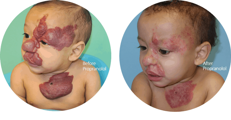
Vascular Birthmarks Conditions: Hemangioma

What is a hemangioma?

Hemangiomas are benign blood vessel tumors that occur in childhood. The most common type is the “infantile hemangioma” version (“strawberry mark”). Children are not born with these tumors typically. They appear in the first few days to weeks of life (can extend up to 6 months of age) as a faint red or pink blemish that can rapidly grow into a larger mass.

(626) 765-6885

The source of hemangioma is yet to be determined, but some research suggests it may be linked to genetics or other diseases.
Hemangiomas range in size from a few millimeters to several centimeters.

Growth will peak at around 12 months of age and plateau thereafter. It will then take several years for the hemangioma to disappear or involute. This takes place usually between 3 and 7 years of age. “Congenital” hemangiomas are hemangiomas that are present at birth. These do not follow the same life cycle as the infantile kind. Rapidly-involuting congenital hemangiomas (RICH) will disappear by 12 months of age, whereas non-involuting congenital hemangiomas (NICH) will persist throughout life.

What causes hemangiomas?

A hemangioma is a mass of extra blood vessels that gather in one area. While the cause of this condition remains unknown, studies suggest it might be a genetic condition or illness. However, we know that it occurs when there’s an unusual concentration of extra blood vessels.

Non-involuting congenital hemangioma of arm.
Non-involuting congenital
hemangioma of arm.

Types of hemangiomas

Hemangiomas come in two main varieties: infantile and congenital.

Infantile hemangiomas

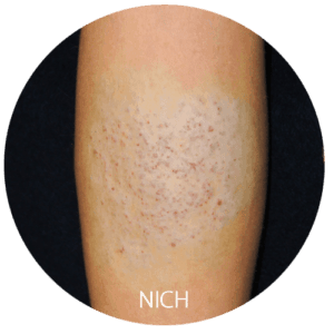
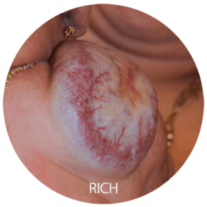
These types of hemangiomas are usually not present at birth, and they start growing after birth. They look red and grow rapidly. Subtypes of infantile hemangiomas are:

  • Superficial Hemangiomas, or “strawberry” hemangiomas, appear as bright red tumors.
  • Deep Hemangiomas can appear under or on the skin that may appear normal or have a bluish hue.
  • Mixed Hemangiomas have both superficial and deep components.
  • Nodular hemangioma is the most common form. They are discreet and oval or round. Besides, these hemangiomas tend not to have a connection with malformations.
  • Large hemangiomas with characteristic geographic shapes that span a wide area of the body can be a sign of greater complications and underlying developmental issues.

Congenital hemangiomas

Prenatal ultrasounds can detect hemangiomas, which are vascular lesions that have developed in the uterus and are present at birth. These lesions usually appear round or oval and may be pink, purple, or a mixture of colors. Additionally, the skin growth is usually raised and warm to the touch.

Hemangioma Diagnosis

A physical examination and review of the patient’s history are typically all that is necessary for diagnosing these vascular malformations. If there is an underlying lesion, an ultrasound or MRI can identify it further.

Hemangiomas may, though rarely, be linked to other syndromes. To determine such a connection, it is possible that further evaluation and testing.

What are the options for hemangioma treatment?

Most hemangiomas, being of the “infantile” variant, require no specific intervention. Over time, the hemangioma will initially increase in size, then plateau, and continue on to involution. Often, the entire hemangioma will disappear at the end of this process with no residual signs that it was ever there.

Sometimes, hemangiomas may require treatment. This is true of large hemangiomas, those that cause visible disfigurement, ones that have failed to involute completely, or those that can obstruct vital functions such as vision or breathing. In these cases, treatment can take the form of medication or surgery.

The majority of infantile hemangiomas in Los Angeles do not require any type of treatment.
Rapidly-convoluting congenital hemangioma of a child’s shoulder.

Treatment is necessary if there are :

  • Ulceration. Hemangiomas can lead to skin breakdown, which is often painful and can cause permanent scarring. Areas like body folds, diaper regions, and lips are most vulnerable to ulceration.
  • Impairment of vital functions. Depending on their size and location, hemangiomas can impact important functions such as sight, breathing, and eating. For instance, hemangiomas in the eyelid can prevent normal eyesight development. A surgeon must address them immediately.
  • Risk of permanent alteration to appearance. If a hemangioma is on the face or in other easily visible places, treatment may be necessary, as it may cause permanent changes to the skin or develop into a fibrofatty mass.

Localized Treatment Options

  • Timolol, a type of topical beta-blocker, can also treat hemangiomas.
  • Although a topical steroid may be helpful, the use of timolol has become more popular than in previous years.
  • Steroid injections into the hemangioma can be a beneficial form of treatment.
  • A topical wound care routine can aid in the treatment of ulcerated hemangiomas.

Systemic Treatment Options

  • Oral beta-blockers are currently the most widely used form of treatment for hemangiomas that require systemic therapy. Propranolol, which has been in use for several years to treat hypertension, has gained FDA approval for the management of hemangiomas. Side effects may include drops in heart rate, blood pressure, and blood sugar; that is why close monitoring by a medical professional is requisite.
  • Although they are not as widely used as oral beta blockers, oral steroids may still be beneficial in certain instances.

Other Treatment Options

  • Laser treatments can be beneficial for treating ulcers in hemangiomas and lessening any remaining redness after involution.
  • When a child is between 3 and 4 years old, a surgical procedure can be a great way to remove and repair any residual tissue that may remain after involution. In certain cases, your surgeon may recommend earlier surgical intervention.
Before and after photos of child treated with propranolol for infantile hemangioma of face and chest.
Rapidly-convoluting congenital hemangioma of a child’s shoulder.

Surgery is only an option if there are hemangiomas that are causing symptoms (eg, ulceration or frequent bleeding) or functional obstruction such as those located over the eyelid or inside the airway. Sometimes, smaller hemangiomas may benefit from early excision to avoid social insecurity during childhood. The decision to pursue a surgical option should be largely guided by the surgeon, who will evaluate whether or not any particular hemangioma can be excised without causing significant secondary deformity.

Interviewed and researched many doctors for my procedure. The difference between Dr. Panossian and the others was he took the time in our pre and post appointment. I did not feel rushed. He treated me as a human being and I felt he was very present in every moment with me and not distracted with other things. Very professional, but approachable and personable.

  • Star full Star full Star full Star full Star full 5 stars
  • T.J.

View all Testimonials

When to see a doctor for hemangioma

The doctor will regularly track the development of your kid’s hemangioma. If the hemangioma bleeds, forms a sore or appears infected, contact their doctor immediately.

If this condition affects your child’s ability to see, breathe, hear, or go to the bathroom, seek medical attention.

Is a hemangioma the same as a port wine stain?

There is a lot of misinformation in the world of vascular birthmarks. A study by our own practice revealed a rate of misdiagnosis of more than 65 percent among patients referred. Hemangiomas are NOT the same as port wine stains. Hemangiomas are benign blood vessel tumors that have a fairly predictable life cycle as described above. Port wine stains are capillary malformations that are present at birth and persist throughout life. The importance of making the correct diagnosis is a crucial factor. Treatment options are vastly different between these two conditions. Port wine stains are largely treated with pulsed-dye lasers. Depending on the size of the stain, several treatments over the course of a lifetime are necessary. Early treatment when the port wine stain is light is now recommended. Although certain hemangiomas may respond well to similar laser treatments, this is still not effective for large ones.

In addition, there are other considerations with port wine stains. Some of these are associated with overgrowth conditions when located over a limb. A child may demonstrate a limb length discrepancy as he or she grows. This is sometimes pronounced during puberty and may require treatment in the form of orthotics if it significantly affects walking. In addition, port wine stains occurring over the face need to be scrutinized closely for associated problems, including glaucoma, migraines, and seizures. The combination of these symptoms is known as Sturge-Weber syndrome. Although rare, it is important to recognize this condition and have a multidisciplinary team in place including a plastic surgeon, neurologist, and ophthalmologist.

Learn more about Hemangioma treatment with Dr. Panossian’s expertise. Schedule a consultation at his office in Pasadena or you can call us at 626-765-6885. Our team is ready to help you.

Why choose Dr Panossian

Last modified by Dr. Andre Panossian

Schedule a
Consultation

"*" indicates required fields

By submitting this form I agree to the Terms of Use
This field is for validation purposes and should be left unchanged.