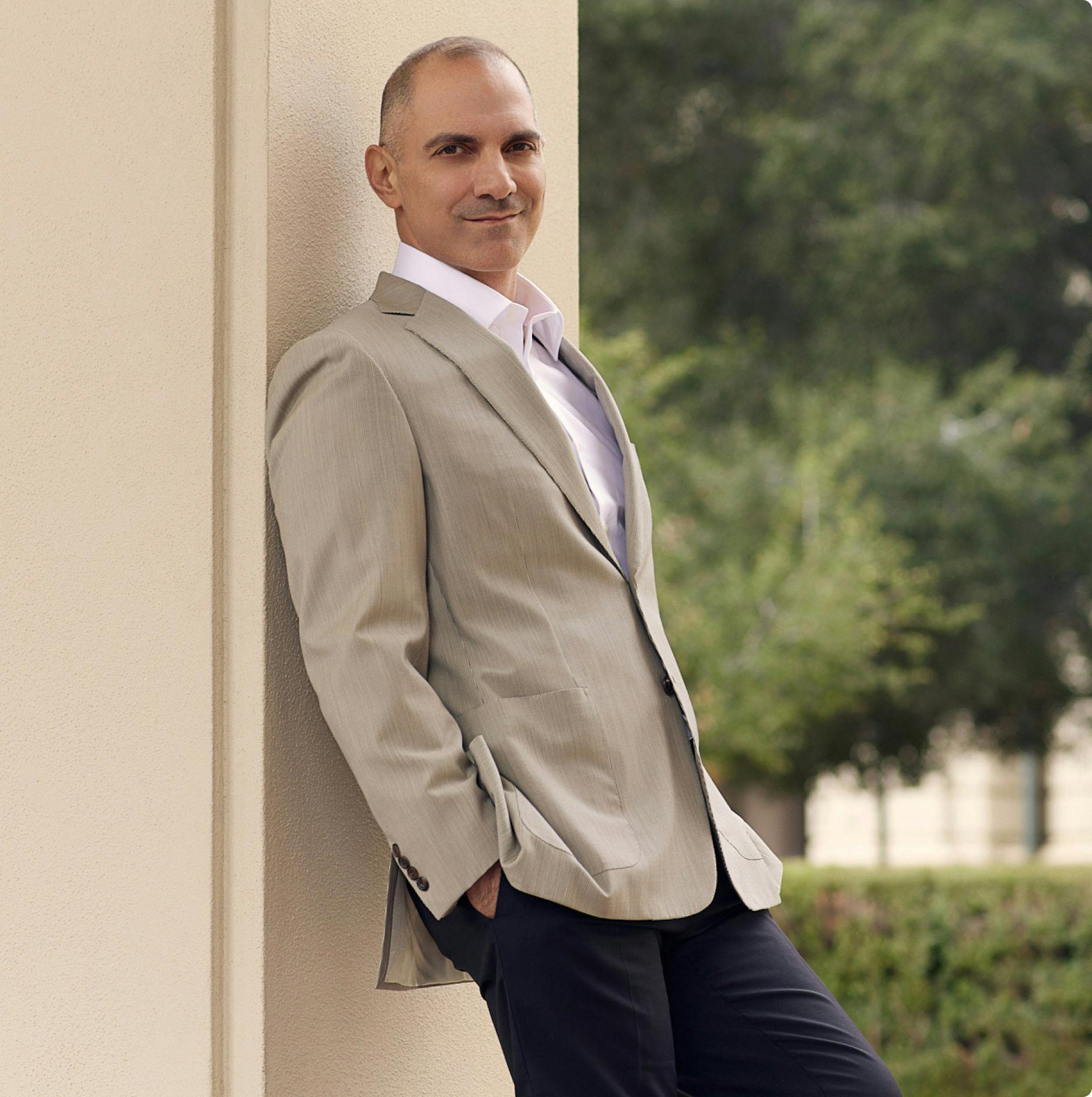
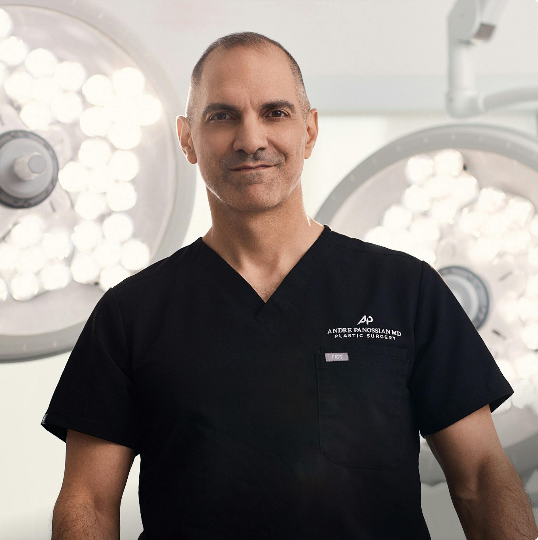
Enhance Ear Form and Function with Dr. Panossian in Pasadena

It can be quite a challenge to watch your child living with microtia, facing daily challenges and emotional insecurities due to the appearance and functional limitations of the ear. Thankfully, Dr. Panossian brings a wealth of reconstructive expertise to microtia surgery, offering personalized solutions to enhance the ear's form and function. With a compassionate approach, Dr. Panossian understands the profound impact that microtia can have on self-esteem and social interactions. As a specialist in reconstructive surgery, trained to address complex craniofacial conditions, Dr. Panossian is dedicated to providing patients in Glendale, Pasadena, and the greater Los Angeles area with transformative results that go beyond cosmetic enhancement, with newfound confidence and hope for the future.

Understanding Microtia

Microtia is a congenital condition characterized by underdeveloped or malformed ears, often present at birth. This rare abnormality affects approximately 1 in 10,000 infants, with varying degrees of severity. Typically, microtia manifests as a small, misshapen external ear lacking the structural support found in fully formed ears. Sometimes, microtia is accompanied by aural atresia, where the ear canal is narrow or absent, leading to hearing impairment.

Anotia denotes the complete absence of an ear, while microtia is classified into different grades based on the extent of ear deformity:

  • Grade I: The ear and ear canal are smaller than average, but external and internal structures are formed.
  • Grade II: The external ear is deformed, and the ear canal may be narrowed or underdeveloped.
  • Grade III: The external ear is more severely misshapen and small and often lacks cartilage. The ear canal may also be narrowed or absent. Grade III represents the most severe form of the condition.
  • Grade IV: Complete absence of the external ear, with the ear canal typically underdeveloped or absent.

The exact causes of microtia are not always known, but they are believed to involve issues with cell development or insufficient blood supply during fetal development. Certain maternal infections, such as rubella, and exposure to certain medications during early pregnancy may also increase the risk of microtia. Medications like retinoic acid, Accutane, clomiphene, and thalidomide have been linked to microtia when used during pregnancy.

Microtia, particularly with hearing loss due to aural atresia, may result in disability, affecting a child's communication skills and learning abilities. Social Security income may help cover some medical expenses associated with microtia-related disabilities. While bone-conduction hearing devices like BAHA (Bone Anchored Hearing Aid) or cochlear implants can help restore hearing, reconstructive surgery aims to improve the ear's form and function.

Banner media
mother holding her toddler son on bed

Ear Reconstruction Surgery

In cases of microtia, where the outer ear is underdeveloped or absent, ear reconstruction surgery offers a transformative solution. This procedure aims to rebuild the ear's structure, restoring both form and function. Dr. Andre Panossian specializes in ear reconstruction, utilizing advanced techniques to create natural-looking ears tailored to each patient's unique anatomy. Ear reconstruction surgery offers numerous benefits, including improved appearance, restored facial symmetry, and enhanced self-confidence. By restoring the external ear's structure, patients can experience a significant improvement in both aesthetic and functional outcomes.

Ear ReconstructionBefore & After

View Gallery

The Surgical Process

  • Initial Evaluation: Patients undergo a comprehensive evaluation with Dr. Panossian before undergoing ear reconstruction surgery. During this consultation, Dr. Panossian assesses the patient's anatomy, discusses their goals and expectations, and recommends the most suitable approach for reconstruction.
  • Surgical Procedure: The procedure for ear reconstruction varies depending on the chosen technique. For rib cartilage grafts, Dr. Panossian carefully harvests cartilage from the ribcage, sculpts it into the desired shape, and implants it into the ear area. Alternatively, MEDPOR implants are customized and inserted into the ear site to create the framework for the new ear.
  • Recovery and Follow-Up: Patients need a recovery period for proper healing following ear reconstruction surgery. Dr. Panossian provides detailed postoperative instructions to help patients navigate the recovery process. Regular follow-up appointments are scheduled to monitor progress and address any concerns.

Surgical Techniques

  • Rib Cartilage Graft: One approach to ear reconstruction involves using rib cartilage from the patient's body. During this procedure, Dr. Panossian carefully harvests cartilage from the ribcage and sculpts it to form the framework of the new ear. This technique allows for precise customization, resulting in an ear closely matching the opposite side's size and shape.
  • MEDPOR Implant: Ear reconstruction can be achieved using a synthetic implant known as MEDPOR. This porous, biocompatible material integrates with the surrounding tissues, providing a durable framework for the new ear. Dr. Panossian meticulously shapes the MEDPOR implant to achieve a natural appearance, ensuring a seamless aesthetic outcome.
Background media

Consultation and Preparation

The journey towards ear reconstruction begins with an initial consultation with Dr. Andre Panossian. During this meeting, Dr. Panossian thoroughly evaluates the patient's condition, considering their medical history, previous treatments, and aesthetic goals. This assessment allows Dr. Panossian to develop a personalized treatment plan tailored to the patient's needs.

Dr. Panossian takes the time to discuss all available options for ear reconstruction, including surgical techniques, potential outcomes, and associated risks. Patients are encouraged to ask questions and express concerns, ensuring they feel confident and informed about their decisions. Throughout the consultation, Dr. Panossian provides realistic expectations regarding the surgical procedure, recovery timeline, and anticipated results. Patients can approach the procedure with confidence and clarity by setting clear expectations.

Before undergoing ear reconstruction surgery, patients undergo a comprehensive medical evaluation to ensure they are suitable candidates. This evaluation may include laboratory tests, imaging studies, and consultations with other medical specialists if necessary.

Dr. Panossian provides detailed preoperative instructions to help patients prepare for surgery. These instructions may include guidelines on medication management, dietary restrictions, and lifestyle modifications leading up to the procedure. Dr. Panossian works closely with the patient's medical team to coordinate all aspects of care leading up to the surgery. Preparation may involve communicating with primary care physicians, anesthesiologists, and other healthcare providers to ensure a seamless and safe experience for the patient.

Ear Reconstruction Recovery

After ear reconstruction surgery, Dr. Panossian's experienced medical team closely monitors patients in the recovery area. Any discomfort or pain is managed with appropriate medications, and patients are given instructions on caring for their surgical sites. Dr. Panossian ensures that patients are stable and comfortable before being discharged home. However, the care continues; patients receive detailed postoperative instructions and are scheduled for follow-up appointments to monitor their progress and address concerns.

Patients receive guidance on caring for their surgical incisions to promote proper healing and minimize the risk of infection. This guidance may include instructions on keeping the surgical sites clean and dry, changing dressings as needed, and avoiding activities that could strain the ears. Dr. Panossian provides recommendations for managing pain and discomfort during the recovery period. Depending on the individual's needs, this may involve over-the-counter pain relievers or prescription medications.

Patients are advised to avoid strenuous activities and heavy lifting for a certain period following surgery to ensure optimal healing. Dr. Panossian provides specific guidelines on when it's safe to resume normal activities based on each patient's recovery progress. Patients are scheduled for follow-up appointments with Dr. Panossian to assess their healing progress and address any concerns or questions they may have. These appointments allow Dr. Panossian to monitor the surgery's results and make any necessary adjustments to the treatment plan.

Even after the initial recovery period, Dr. Panossian continues to monitor patients' progress to ensure the best possible outcomes. Regular check-ups are scheduled to assess the long-term stability and function of the reconstructed ears and address any issues that may arise over time.

Schedule A Consultation

Book Now
Contact us media

What to Expect: The Cleft Lip & Palate Surgery

Cleft lip and palate repair surgeries are typically performed under general anesthesia and may require an overnight hospital stay for observation, particularly in the case of palate repair. Lip repair involves moving adjacent segments of the upper lip together to re-establish continuity and function. Repairing the underlying muscle sphincter (orbicularis oris) is vital to restoring proper upper lip function. Palate repair involves repairing the hard and soft palate layers separately, ensuring proper soft palate repair, which is critical for speech development. Additional procedures may be performed as needed, such as bone grafting for gum line clefts or secondary speech-improvement surgeries. Surgical times vary depending on the complexity and extent of the procedure but generally range from 2 to 3 hours. Some surgeries may involve combination procedures, such as ear examinations under anesthesia or bone grafting. Recovery from surgery involves close monitoring for bleeding or respiratory difficulties, along with a soft diet and restrictions on physical activity as appropriate. Postoperative follow-up visits are typically scheduled to assess healing progress and address concerns. Surgical or secondary procedures may be considered based on individual needs and outcomes.

Cleft Lip & Palate Surgery Recovery

Following cleft lip repair, many centers now practice outpatient procedures. The incision is typically sealed with skin glue or small sutures, which may require removal after one week. Children can resume feeding immediately after surgery and return to their usual level of physical activity, supervised but without restraints, as infants typically do not feel the urge to manipulate the surgical site. Parental involvement is crucial, and a "less-is-more" approach is often favored. Follow-up appointments are scheduled at 1 and 4 weeks postoperatively, during which scar management techniques such as massage, taping, sunblock application, and moisturizing cream usage may be introduced.

In contrast, cleft palate repair usually necessitates overnight hospitalization to monitor potential bleeding or respiratory complications. The child's ability to maintain oral hydration and feeding is closely observed, with some expected oral and nasal bleeding for up to one week postoperatively. Follow-up appointments occur at 1 and 6 weeks postoperatively, during which a soft diet is mandated while the child is allowed to resume regular physical activity immediately after surgery.

Speech therapy is integral to cleft palate management, as approximately two-thirds of patients may experience speech disturbances, often characterized by excessive nasal air escape or hypernasality. Early intervention is key to preventing the formation of improper speech habits that can exacerbate long-term speech difficulties. While about one-third of patients may achieve improvement with long-term speech therapy alone, another third may require surgical intervention to enhance palatal movement efficiency and reduce hypernasality. This surgical intervention, performed by Dr. Panossian, aims to make speech more intelligible and can significantly improve long-term outcomes for patients with cleft lip and palate.

Background media

Risks and Potential Complications of Cleft Lip & Palate Surgery

While cleft lip and palate surgeries have undergone extensive refinement over decades, like any surgical procedure, they carry inherent risks and potential complications. These include the possibility of bleeding, infection, or anesthesia-related issues, albeit rare. For cleft lip repair, there's a slight risk of undesirable scarring, such as hypertrophic scarring or keloids, along with the potential for residual deformities like excessive upper lip bulk or asymmetry. In rare cases, wound dehiscence, or separation, may occur post-surgery, necessitating possible revision surgery for correction.

In cleft palate repair, there's a risk of incision line breakdown, with potential communication between the oral and nasal cavities, known as oronasal fistula, which may require repair if significant or affecting speech. Soft palate repair dehiscence could impair palatal muscle function, necessitating corrective measures. While extremely rare, postoperative airway swelling leading to respiratory difficulties may occur. Additionally, persistent speech issues are common, underscoring the importance of early intervention with a speech therapist.

Secondary procedures for speech correction, such as velopharyngeal insufficiency (VPI) treatment, carry additional risks, including respiratory compromise, albeit rare. There's also a possibility of exacerbating sleeping difficulties like obstructive apnea or snoring in some cases. Despite efforts to improve speech, procedures may not always fully address speech concerns, leading to ongoing reliance on speech therapy for long-term management.

Doctor media
Doctor media
Doctor media

Why Choose Dr. Panossian?

Choosing Dr. Panossian for your ear reconstruction surgery means entrusting your care to a highly skilled and compassionate expert. With extensive training in reconstructive surgery and a proven track record of excellence, Dr. Panossian is equipped to address the unique challenges of microtia with precision and care. His expertise in groundbreaking procedures, such as leading Haiti's first-ever conjoined twin separation, showcases his proficiency in complex reconstructive surgeries. Dr. Panossian understands the emotional impact of microtia and is dedicated to providing personalized care that meets each patient’s needs. By reshaping and enhancing the appearance of the ear, Dr. Panossian not only restores physical form but also offers a renewed sense of confidence and self-assurance. If you’re seeking ear reconstruction in Glendale, Pasadena, or the greater Los Angeles area, schedule your consultation with Dr. Panossian today.

Microtia FAQ

What factors influence the cost of ear reconstruction surgery?

Does insurance typically cover ear reconstruction surgery?

Can accessory auricles be addressed during ear reconstruction surgery?

How are different ear shapes created based on the patient's anatomy?

What procedures does Dr. Panossian specialize in?

Does microtia affect intellectual abilities?

Can microtia be detected during ultrasound scans?

What factors influence the cost of ear reconstruction surgery?

The cost of ear reconstruction surgery can vary significantly depending on insurance coverage, whether an in-network provider performs the procedure, the type of procedure involved, the need for ear canal reconstruction, and the installation of a cochlear implant. The case's complexity will also impact the total cost, particularly in multi-step rib cartilage reconstructions. Patients can request estimates from Dr. Panossian's staff after consulting with their insurance company.

Does insurance typically cover ear reconstruction surgery?

In most cases, insurance companies do not cover earlobe reconstruction surgery as it is considered cosmetic and not essential for hearing function. Earlobe surgeries are often sought for cosmetic reasons related to aging, piercings, or regrettable alterations such as stretched earlobes. Costs for earlobe reconstruction surgery are relatively low per ear, typically ranging from $400 to $1,200.

Can accessory auricles be addressed during ear reconstruction surgery?

Accessory auricles, or abnormal growths on the ear, can be removed during cosmetic ear surgeries through excision or laser treatment. While insurance generally does not cover elective cosmetic procedures, removing accessory auricles is often straightforward and can be performed as part of ear reconstruction surgery.

How are different ear shapes created based on the patient's anatomy?

The pinna, or outer ear structure, varies uniquely for each individual. When reconstructing the ear surgically, Dr. Panossian utilizes either the patient's cartilage or MEDPOR porous polyethylene, skillfully shaping them with specialized surgical tools. In microtia cases where one ear is affected, and the other remains normal, Dr. Panossian can use the unaffected ear as a reference to create a closely matched replica. It's important to note that pediatric and adult ear sizes may require different surgical techniques to achieve optimal results.

What procedures does Dr. Panossian specialize in?

Dr. Panossian specializes in plastic surgery of the face and body, focusing on craniofacial and reconstructive surgery. He has extensive experience in operating on children with various deformities and neurological issues, including facial paralysis and neurofibromatosis. Dr. Panossian is dedicated to enhancing irregular anatomy to improve the quality of life for his pediatric patients.

Does microtia affect intellectual abilities?

Microtia, an underdeveloped ear condition, does not directly impact a child's cognitive abilities. However, hearing loss associated with microtia can affect learning in environments reliant on hearing. Parents should seek early intervention by scheduling appointments with medical specialists to address the child's hearing needs. Hearing aids and American Sign Language can aid in communication skills development for hearing-impaired infants.

Can microtia be detected during ultrasound scans?

Microtia, a congenital abnormality, is typically not detectable during prenatal ultrasound scans. While advanced imaging techniques may be used, detecting ear abnormalities is often not feasible before birth. Early diagnosis focuses more on organ and limb development, and interventions for microtia cannot begin until after birth. Following obstetrician medical advice and avoiding medications linked to microtia is essential to minimize associated risks.

Contact us media Contact us media

© Andre Panossian, M.D.. All Rights Reserved.

Accessibility: If you are vision-impaired or have some other impairment covered by the Americans with Disabilities Act or a similar law, and you wish to discuss potential accommodations related to using this website, please contact our Accessibility Manager at 626-385-4659.
Contact Us